Home
/ Back Of Skull Anatomy Labeled : The Base of the Skull. Inferior view - #anatomy images ... / The skull or known as the cranium in the medical world is a bone structure of the head.
Back Of Skull Anatomy Labeled : The Base of the Skull. Inferior view - #anatomy images ... / The skull or known as the cranium in the medical world is a bone structure of the head.
Back Of Skull Anatomy Labeled : The Base of the Skull. Inferior view - #anatomy images ... / The skull or known as the cranium in the medical world is a bone structure of the head.. Skull, parietal bone, nasion, frontal process, lacrimal bone, nasal bone, spehenoid lesser wing, middle nasal. We use cookies to ensure that we give you the best experience on our website. Anatomy visible in the medical illustration includes: Last updated on fri, 26 feb 2021 | human anatomy. The frontal, parietal, temporal and occipital bones are joined at the cranial sutures.
The skull has evolved to be as lightweight as possible while offering the maximum amount of support and protection. Excluding ear ossicles, it is made of 22 bones. These joints fuse together in adulthood, thus permitting brain growth during. It is comprised of many bones, formed by intramembranous ossification, which are joined together by sutures (fibrous joints). Human skull anatomy, the skeletal structure of the highest point of vertebrates, made out of bones or ligament, which shapes a unit that, ensures the brain and some vibe organs.
Beauchene Skull Model - HUMAN ANATOMY WEB SITE from mesa-anatomy.weebly.com Related posts of bone of back of skull. When this deck is imported into the desktop program, cards will appear as the deck author has made them. They don't move and united into a single unit. In order to be light, the skull is made up by flat and irregular bones, and has hollow spaces called the sinuses. All the bones of skull, joined together by sutures… the skull is subdivided into 2 parts: Learn more about the anatomy and function of the skull in humans and other vertebrates. Please feel free to download and print. Skull, skeletal framework of the head of vertebrates, composed of bones or cartilage, which form a unit that protects the brain and some sense organs.
Please feel free to download and print.
Which bone (yellow) is centrally located and joins with most. 11.3 axial muscles of the head, neck, and back. Skull anatomy gross anatomy brain anatomy medical anatomy anatomy study physician assistant education al dente. It supports and protects the face and the brain. This is a model of the human (homo sapiens) skull. The skull is the bony skeleton of the head. Human anatomy for muscle, reproductive, and skeleton. Foundational anatomy provides medical students with the necessary background in anatomy for success in clerkships. It offers protection to the brain, eye balls, inner ears, and nasal passages. We also cover the ear bones and the hyoid bone.transcript/notesskull. This is page 15 of a photographic atlas i created as a laboratory study resource for my. The skull performs vital functions. Adelstein on skull labeling anatomy:
Learn more here you are seeing a 360° image instead. Related posts of bone of back of skull. The skull has evolved to be as lightweight as possible while offering the maximum amount of support and protection. It offers protection to the brain, eye balls, inner ears, and nasal passages. Skull, skeletal framework of the head of vertebrates, composed of bones or cartilage, which form a unit that protects the brain and some sense organs.
Skeletal System Diagrams from jb004.k12.sd.us Anatomical structures of the skull include: Skull, parietal bone, nasion, frontal process, lacrimal bone, nasal bone, spehenoid lesser wing, middle nasal. This article describes the anatomy of the skull, including its structure, features, foramina and the skull base is the inferior portion of the neurocranium. This is a model of the human (homo sapiens) skull. It was then cleaned, adapted and polypainted this model is part of a comparison with the skull of a human. Related posts of bone of back of skull. 11.3 axial muscles of the head, neck, and back. All the bones of skull, joined together by sutures… the skull is subdivided into 2 parts:
It was then cleaned, adapted and polypainted this model is part of a comparison with the skull of a human.
Labelled poster sized anatomical illustration of the bones of the skull in anterior view available to license on a rights managed basis. Which bone (yellow) is centrally located and joins with most. That is how the doctor insights on: Anatomy and physiology7.2 the skull. Examine the cranial bones of the articulated human skull and the sectioned skull. Please feel free to download and print. It is comprised of many bones, formed by intramembranous ossification, which are joined together by sutures (fibrous joints). Learn more here you are seeing a 360° image instead. Review a textbook section on the skull. Human anatomy for muscle, reproductive, and skeleton. The major sutures are the coronal suture, sagittal suture, lambdoid suture and squamosal sutures. The skull includes the upper jaw and the cranium. Adelstein on skull labeling anatomy:
This is a model of the human (homo sapiens) skull. Please feel free to download and print. Human skull anatomy, the skeletal structure of the highest point of vertebrates, made out of bones or ligament, which shapes a unit that, ensures the brain and some vibe organs. In this video we discuss the locations of the bones of the skull and label them. Review a textbook section on the skull.
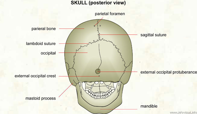
A&P: Chapter 7 The Skull at Carolinas College of Health ... from classconnection.s3.amazonaws.com The major sutures are the coronal suture, sagittal suture, lambdoid suture and squamosal sutures. At the same time the bones grow larger by growing back into the growth plates. The skull is the bony skeleton of the head. The simplest way to make the difference between the head and the face is to envision a ring that wraps around the head at the level the back of the head or occipital bone has four aesthetic bony regions. Helpful, trusted answers from doctors: Exterior skull anatomy 3d model. Human skull anatomy, the skeletal structure of the highest point of vertebrates, made out of bones or ligament, which shapes a unit that, ensures the brain and some vibe organs. If you'd like to customize what appears on the front and back of a card, you.
Related posts of bone of back of skull.
Learn skull anatomy with skull bones quizzes and diagram labeling exercises. As a review activity, label figures 13.1, 13.2, 13 3, 13.4, and 13.5. Skull, skeletal framework of the head of vertebrates, composed of bones or cartilage, which form a unit that protects the brain and some sense organs. We also cover the ear bones and the hyoid bone.transcript/notesskull. We use cookies to ensure that we give you the best experience on our website. Foundational anatomy provides medical students with the necessary background in anatomy for success in clerkships. The frontal, parietal, temporal and occipital bones are joined at the cranial sutures. It was then cleaned, adapted and polypainted this model is part of a comparison with the skull of a human. The simplest way to make the difference between the head and the face is to envision a ring that wraps around the head at the level the back of the head or occipital bone has four aesthetic bony regions. Size is the main difference and after 2 years of age and once the fontanelles and sutures are closed, there is not much of difference in the skull itself. The major sutures are the coronal suture, sagittal suture, lambdoid suture and squamosal sutures. Review a textbook section on the skull. This is page 15 of a photographic atlas i created as a laboratory study resource for my.
Please feel free to download and print back of skull anatomy. 11.3 axial muscles of the head, neck, and back.

